Farrier + Vet Collaboration
𝐖𝐡𝐞𝐧 𝐅𝐚𝐫𝐫𝐢𝐞𝐫 𝐚𝐧𝐝 𝐕𝐞𝐭𝐞𝐫𝐢𝐧𝐚𝐫𝐢𝐚𝐧 𝐖𝐨𝐫𝐤 𝐓𝐨𝐠𝐞𝐭𝐡𝐞𝐫
A horse’s hoof is a complex structure, and what we see on the outside does not always tell the full story. Collaboration between your veterinarian and farrier can provide valuable insight into the internal structures of the foot and help guide more informed trimming and shoeing decisions.
𝐖𝐡𝐲 𝐌𝐢𝐠𝐡𝐭 𝐑𝐚𝐝𝐢𝐨𝐠𝐫𝐚𝐩𝐡𝐬 𝐁𝐞 𝐑𝐞𝐜𝐨𝐦𝐦𝐞𝐧𝐝𝐞𝐝?
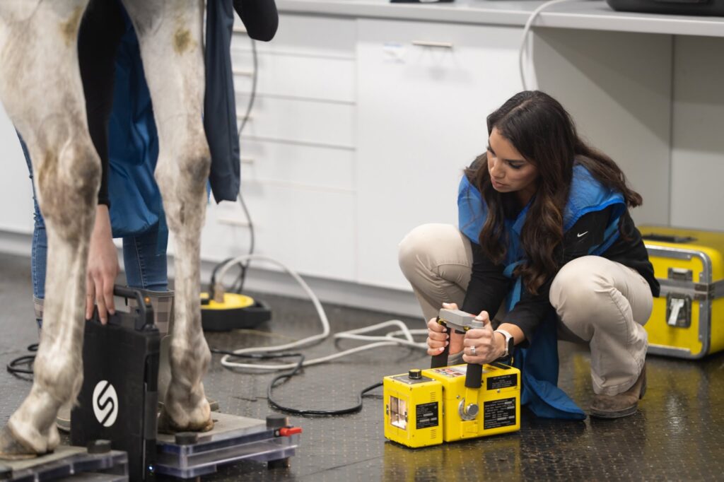
Radiographs can provide an inside view of the hoof and help evaluate:
• Coffin bone position and alignment
• Hoof-pastern axis and overall angles
• Sole depth and protection
• Coffin joint space
• Changes, rotation or sinking of the coffin bone
• Bone remodeling or arthritic changes
• Previous or current hoof-related injuries
• Changes associated with laminitis or other hoof disease
This information can be especially valuable when managing chronic lameness, laminitis, thin soles, abnormal hoof conformation, recurrent hoof problems or a horse that is not responding as expected to routine farrier care.
𝐓𝐡𝐞 𝐁𝐞𝐧𝐞𝐟𝐢𝐭 𝐨𝐟 𝐂𝐨𝐥𝐥𝐚𝐛𝐨𝐫𝐚𝐭𝐢𝐨𝐧
Radiographs allow the veterinarian to evaluate the internal structures while the farrier can use that information to make precise adjustments to the external hoof. When both professionals communicate and work from the same information, trimming and shoeing decisions can be better tailored to the individual horse.
For the horse owner, this means a more complete understanding of what is happening inside the hoof. For the veterinarian, it provides an opportunity to incorporate the farrier’s knowledge of the horse’s hoof history, conformation and response to previous shoeing. For the farrier, it provides valuable information that cannot be determined from the outside alone.