Arthritis in Horses

What is arthritis?
Arthritis is an inflammation of a joint or joints that causes pain and stiffness but the word ‘arthritis’ is often used to cover a range of conditions, many of which are not true arthritis. Related conditions including degenerative joint disease (DJD), osteoarthritis (inflammation involving bone changes), synovitis (inflammation of the synovium, i.e., joint lining), are often imprecisely called ‘arthritis’.
How does arthritis occur?
Normal, painless joint function is essential for an athletic animal and for this to occur, each component of the joint structure must be healthy and working properly. Smooth movement of the opposing bone ends relative to each other and the surrounding structures is achieved by its complex structure: the special shape of the end of each bone, the covering of smooth cartilage over the end of each bone, the presence of synovial fluid (‘joint oil’) that lubricates the cartilage, all contained within the joint capsule that forms an elastic pouch that encloses the structures of the joint, and in some cases contains supporting ligaments.
Arthritis often develops following interference with normal structure and function. Damage to the cartilage and/or the bone through trauma (injury) or infection results in roughening of the smooth surfaces. Movement of the roughened bone ends and/or damaged cartilage results in inflammation, that produces mediator chemicals that damage the synovial fluid, all of which causes swelling of the joint, pain and restriction of movement. This initial response is called acute arthritis. Injury to the joint capsule and its ligaments can also trigger inflammation resulting in thickening of the capsule, reducing its elasticity and thinning of the joint fluid, reducing its lubricating ability.
Over time, new bone is formed in response to surface damage. This new bone is rough and is not covered with protective cartilage, therefore interferes with joint movement and causes pain. These longer-term effects are called chronic arthritis.
Degenerative joint disease can result in the same long-term changes but in contrast to true arthritis, is usually not associated with pain or inflammation in the early stages. In DJD the joint structures respond to wear and tear by gradually changing shape and elasticity. Many horses with DJD move soundly. In others, these changes progress to a stage where the horse goes lame.
What are the signs of arthritis in horses?
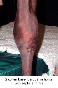
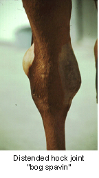
Joint inflammation increases the amount of fluid in the joint and this usually causes visible and/or palpable bulging of the joint capsule (e.g., ‘bog’ spavin, ‘popped’ knee, ‘windgall’). However, not all joint swellings are due to arthritis. For example, a simple ‘strain’ may cause transient joint swelling. In arthritis, there is pain when the affected joint is flexed (bent) and the horse may be lame or stiff at the walk or trot. In acute arthritis, the swollen joint may appear warm to touch.
In acute arthritis caused by infection (‘septic’ arthritis) there is usually severe inflammation, pain and lameness. If not quickly controlled, this condition can cause severe destruction of the joint surfaces that may end a horse’s athletic career and may even necessitate euthanasia on humane grounds. Septic arthritis must therefore be treated as a medical emergency. Infection may be introduced directly by injury (joint puncture) or via the bloodstream from a primary focus of infection elsewhere.
With DJD and chronic arthritis, joint mobility is reduced but there may not be pain on flexion of the joint. There may be lameness that has gradually worsened over time or lameness that improves with exercise, i.e., the horse ‘warms up’.
In cases where there are multiple joints involved, the horse may appear generally stiff at one or all gaits. In an older horse, the main sign of abnormality may be difficulty in standing up after a period of lying down.
How is the diagnosis of arthritis confirmed?
If you are worried that your horse might have arthritis, you should ask your veterinarian to examine him. This will involve feeling the limbs to look for abnormalities such as swelling, reduction in range of movement or pain on flexion. In acute arthritis, the joint may be hot and swollen when examining for lameness or stiffness. This might mean just walking and trotting in hand if the lameness is very obvious but if the gait abnormality is only subtle, the horse may need to be ridden or lunged.
In many cases, it is not possible to confirm the diagnosis or its significance by clinical examination alone. Further examinations may involve nerve block examinations, where local anesthetic is injected around specific nerves to abolish pain from the parts of the leg that these nerves supply, confirming the site of the pain radiographic (x-ray) examinations to rule out fractures and to look at the bones at the joint surfaces for signs of injury, degeneration or abnormality. Radiographs are not able to demonstrate cartilage damage so are more helpful for chronic arthritis and DJD cases, where the new bone formation can be demonstrated, magnetic resonance imagining (MRI) may help to diagnose cartilage and other soft tissue joint injuries or abnormalities. This technique is currently very expensive. Joint fluid collection for laboratory analysis to look specifically for signs of infection maybe helpful. Nuclear bone scan may be helpful in complex cases to image a specific area of increased bone metabolism. This requires the injection of a radioactive bone tracer, with a short half-life, and imaging with a gamma camera.
How is arthritis in horses treated?
Some horses with arthritis respond simply to rest for a period of weeks to months. In most cases this will mean that the horse is confined to a stable. Do not be tempted to turn the horse out during this time. Even though you are not riding him, he could still gallop or leap about and cause more damage to the inflamed structures.
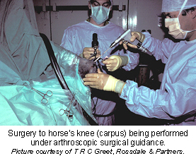
In other cases, the condition will require treatment with non-steroidal anti-inflammatory drugs such as phenylbutazone or meclofenamic acid, that reduce pain and inflammation and help the joint to return to normal function. In some cases of arthritis it may be necessary or useful to inject medication into the joint itself. The more commonly used intra-articular preparations are corticosteroids and hyaluronic acid (artificial joint fluid). These reduce inflammation within the joint and help re-establish the normal lubricating properties of the joint fluid. While this treatment is often initially effective, some cases require repeated injections. In some cases, for example, where there has been extensive new bone formation or where a chip fracture has occurred as a consequence of DJD, arthroscopic surgery may be helpful to give the joint a better chance for recovery. The choice of appropriate therapy must be based upon an accurate and complete diagnosis and the particular needs of the individual case. Your veterinarian will help you with this.
If, in spite of all efforts, severe chronic arthritis remains and the horse has a persistently painful joint with little movement, it is sometimes possible to restore soundness by surgically fusing the affected joint (arthrodesis). This is occasionally used for the pastern and lower hock joints but only as a last resort after all other forms of treatment have been tried.
What is the prognosis?
If acute arthritis is diagnosed and successfully treated early, a complete cure may occur, leaving no residual abnormality. If the inflammation does not respond to treatment and/or is complicated by infection or cartilage or joint injury, new bone may form and the joint may be permanently affected by chronic arthritis. If controllable by careful management and appropriate treatments, low-grade uncomplicated arthritis may not cause lameness with associated pain and restricted mobility.
Caution
It is important to realize that a horse can have arthritic changes that are visible on radiographs but may still move soundly and able to compete successfully. Also, it is possible for a horse to suffer from arthritis, involving the cartilage and joint fluid that will not produce demonstrable radiographic changes. Both scenarios can produce difficulties when a horse is being examined for purchase, i.e., ‘vetted’. If radiographs are taken as a requested routine part of a pre-purchase examination, the images must be carefully interpreted with reference to the findings of the clinical examination.
Contributors: Deidre M. Carson, BVSc, MRCVS & Sidney W. Ricketts, LVO, BSc, BVSc, DESM, DipECEIM, FRCPath, FRCVS
Edited by Kim McGurrin BSc DVM DVSc Diplomate ACVIM
© Copyright 2010 Lifelearn Inc. Used and/or modified with permission under license.


